Noninvasive cardiovascular imaging benefits from MRI and CT scan exploration.
MRI is a technique used to create image slices of the human body that passes radiofrequency waves through a magnetic field to make the hydrogen atoms in body tissues vibrate. It does not use X-rays. Heart MRI enables dynamic visualization of the myocardium and the heart chambers, and thus provides anatomical (heart mass, myocardial thickness, heart valves, pericardium) and functional (volume measurement, ejection fraction measurement, heart component mobility study) information. Injecting gadolinium provides information on the quality of myocardial perfusion and enables the detection of tissue reorganization, particularly ischemia and inflammation. This exam is synchronized with the heart rhythm and requires participation in the form of repeatedly holding your breath for the short period. In some specific cases, analysis of the aorta can also be performed during this exam. Vascular MRI, which is useful for analyzing large and small vessels (aorta, supra-aortic arterial trunks, abdominal arteries, iliac arteries and the lower limbs) is a separate exam. The exam can take from 20 to 40 minutes depending on the indications, and you will be informed ahead of time of the expected duration. There are some contraindications to MRI, which is why you will be given an information sheet before the exam is performed.
Contraindications:
- Pacemaker, implantable automatic defibrillator
- Some older metal heart valves
- Some neurosurgical vascular clips
- Intra-orbital foreign bodies
- Cochlear implants
- Pregnancy
Warnings:
- Let us know if you are claustrophobic (visiting the facility ahead of time can help you deal with feelings of anxiety)
- We may need to test your creatinine levels and test its clearance depending on your age and/or your personal or family history.
To prepare yourself ahead of time, you can download the form from our website.
Why do a stress MRI?
In order to function, the heart muscle receives oxygenated blood from arteries, specifically the coronary arteries. If one or more of these arteries are narrowed due to the presence of artheromatous plaques, this makes it harder for the blood to flow, although enough blood flow may get through to ensure adequate function while at rest. However, when effort is exerted, blood flow to the cardiac muscle through the narrowed artery or arteries may not be adequate. Dipyridamole MRI assesses perfusion of the cardiac muscles by reproducing effects similar to those that occur during exertion (dilation of the coronary arteries) due to injection of a medication, dipyridamole. This exam can detect the presence of coronary artery narrowing with decreased blood flow, which does not manifest as clinical signs. It also looks for any possible scarring from heart infarction.
Before the exam:
- MRI contraindications are eliminated
- You have a pacemaker, a cardiac defibrillator or a prosthetic heart valve (Starr type)
- You have a magnetic cochlear implant, a neurostimulator or a bone growth stimulator
- You have a vascular access port for automated injection of medication (for example, an insulin pump)
- You have iron-containing elements close to your eyes or head (metal filings or clips) or any metallic element in your body
- You cannot undergo MRI because these are contraindications for its use
- If you have asthma, dipyridamole injection is contraindicated
Your information will be sent by the prescribing doctor to the doctor performing the exam. A decision will be made regarding the medications that can be taken before the exam, and you may specifically need to temporarily suspend treatment with beta-blockers (Tenormin, Seloken, Sectral, Lopressor,etc.) the morning of the exam, as they lower the heart rate. (Do not take them in the morning, but bring them to the exam, as you can take them immediately after.)
Important: Do not consume the following foods or beverages in the 24 hours leading up to the exam, as they interfere with dipyridamole and could generate misleading results: coffee, tea, cola drinks, chicory.
Your doctor has suggested that you undergo a cardiac CT. This exam enables analysis of visual slices of your heart, as well as your coronary arteries. This exam enables noninvasive analysis of the anatomy of the coronary arteries, as an alternative to invasive coronary angiography or in addition to other cardiac exams such as a stress ECG or myocardial scintigraphy, if they are inconclusive.
American recommendations and those from the French Society of Cardiology (Société Française de Cardiologie) have permitted its use since 2009. The coronary arteries are very small, between 2 and 4 mm, and run over the surface of the heart, forming a sort of crown ("corona" in Latin) around it. The multi-slice CT scan that we use has a spatial resolution of between 200 and 300 microns, which enables analysis of these small arteries that are not visible by echocardiogram or scintigraphy. The scanner uses X-rays, but at a low dose that will be noted in your report. Nevertheless, this procedure is not recommended for pregnant women. This exam is usually quick (approximately 5 minutes), but your cooperation is important. You must try to remain still and to hold your breath for a few seconds. The exam requires intravenous injection of an iodinated contrast solution, most often through the elbow crease. In the vast majority of cases you will not experience any pain, but will feel a sensation of diffuse warmth when the injection is administered. In rare cases there may be an allergic reaction to the contrast product that is used. If you are aware of having an “iodine allergy”, it is important to let us know before the exam, as well as any history of renal insufficiency. Finally, intravenous injection of a beta-blocker is often needed to slow down the heart. Therefore, we also ask that you notify us if this medication is contraindicated for you (particularly in the case of asthma). Your heart will then be examined for 20 to 30 minutes by your cardiologist, and a report will be given to you at the end of the exam, as well as an explanation of the results.
Our cardiologists are experienced in cardiac imaging and have authored many scientific publications in French and international cardiology journals, include several on controlling X-ray doses.
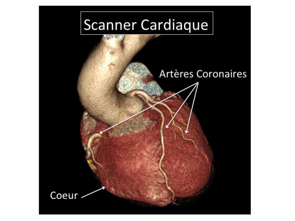
The figure shows a 3D reconstruction of a heart on a CT scan and the coronary arteries on its surface.
Be prepared with the following on the day of the exam:
- The vial or vials or contrast products
- The prescription from your prescribing doctor
- Your previous heart exams
- Valid health insurance card +/- proof of health insurance
- All other certificates: CMU, AME, proof of work accident
- In the case of renal insufficiency: the results of the most recent blood draw
List of doctors in this specialty
-

Dr. PHILIPPE ALLOUCH
Cardiologist
-
Dr. CHRISTOPHE BENSOUDA
Cardiologist
-
DR. LÉA CACOUB
Cardiologist
-
Dr. ARTHUR CESCAU
Cardiologist
-

Dr. RAMI EL MAHMOUD
Cardiologist
-
Dr LAURENT MACRON
Cardiologist
-
Dr. SERGE MAKOWSKI
Cardiologist
-
Dr. JULIEN PASCAL
Cardiologist
-

Dr. DAVID PESENTI-ROSSI
Cardiologist
-

Dr. JULIEN ROSENCHER
Cardiologist