This department oversees care for all emergency or scheduled arterial and venous vascular pathologies. Its medical team combines surgical and radiological knowledge, enabling us to offer a comprehensive range of techniques: conventional, minimally invasive, hybrid and endovascular surgeries. For optimal efficacy, we work in close collaboration with the functional exploration (vascular Doppler), cardiology and heart surgery (combined surgery) teams. We maintain a strong relationship with the nephrology (and its dialysis unit), medical imaging (CT scanner and MRI) and anesthesia-resuscitation teams. The combination of these skills enables us to explore and treat a large variety of vascular pathologies:

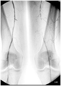
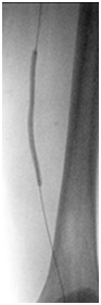
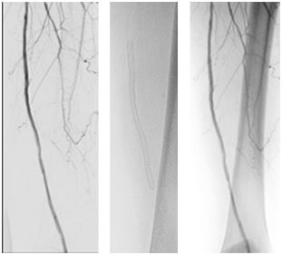
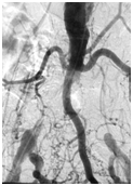
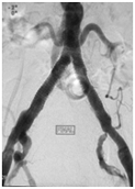
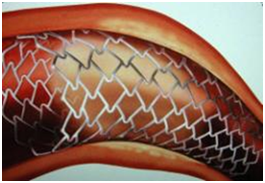
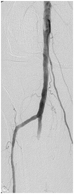
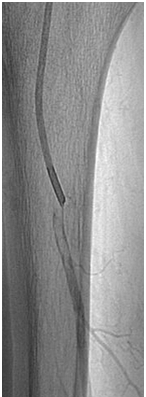
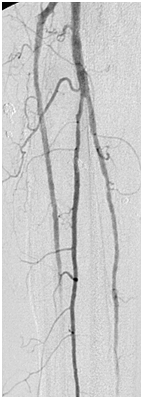
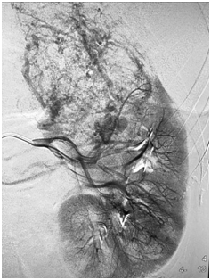
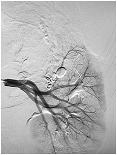
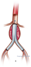
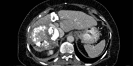
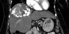
  • Arterial: obliterating arteriopathy of the lower limbs, diabetic arteritis, abdominal aortic or thoracic aneurysms, renal artery stenoses, neck vein stenoses (carotids, subclavian and vertebral).
  • Venous: venous insufficiency (varicose veins treated surgically or by radiofrequency), pelvic varices, etc.
  • Hemodialysis accesses: creation and modification of accesses, phlebography of the extremities, dialysis catheter placement, etc.
  • Pelvis: embolization of uterine fibromas, varioceles, etc.
  • Oncological: chemo-embolization, intra-hepatic catheter placement: files reviewed as part of an oncology multidisciplinary team meeting (MTM) with the 3C CCConcorde team

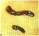
We are available 24 hours a day and 7 days a week to treat emergency cases: acute limb ischemia, embolizations in the event of bleeding, aortic pathologies (aneurysms or dissections), etc.

Resources available:

  • Outpatient and inpatient facilities
  • A dedicated angiography room within the cardiovascular department
  • Surgical suites equipped with angiography machines

Vascular surgery:

Interventional vascular radiology:

Examples of procedures

Treatment of lesions (stenoses and occlusions) of the superficial femoral artery

Leg pain when walking (claudication when walking up to 50 meters). Arteriography confirms occlusion of the femoral artery at the thigh level. Intervention is performed under local anesthesia.

Initial appearance

sans-titre-1

Balloon angioplasty

sans-titre-2

Final result after having inserted a flexible stent

sans-titre-3

Unblocking occlusion of the right and left common iliac arteries

Bilateral pain in the lower limbs when walking that has worsened over several weeks and has not responded to medical treatment.

Before  treatment

sans-titre-4

After revascularization

sans-titre-5

This intervention required the placement of stents in the groin creases using two guide wires under local anesthesia.

sans-titre-6 Several types of stents are available for use in the peripheral arteries. These are small springs that match the diameter of the artery requiring treatment.

Unblocking acute popliteal artery occlusion:

Sudden pain in the leg, which becomes cold, with difficulty moving the toes, associated with a blood clot blocking the popliteal artery (knee).

Emergency removal of the blockage by aspirating the blood clot from vein puncture in the groin crease, performed after confirming the occlusion by Doppler ultrasound.

Initial

sans-titre-7

Aspiration of the clot

sans-titre-8

Final

sans-titre-9

sans-titre-10 Clot extracted by the aspiration probe

Embolization of a benign kidney tumor (angiomyolipoma)

Discovery of a large mass at the upper end of the left kidney. Due to surgical risks, the decision was made to proceed with selective destruction of the tumor by occlusion (embolization) of the vessels supplying it with blood.

sans-titre-11 sans-titre-12

Blood vessel embolization can be achieved using several different materials:

Microparticles with a diameter of 300 to 900 µ

sans-titre-13

Coils (small metal springs)

sans-titre-14

Glue and derivatives (onyx)

sans-titre-15

Maintaining the permeability of an access in a patient on hemodialysis is achieved in collaboration with the team from the hemodialysis department. The exploration (fistulography) is carried out by the vascular radiology service. If treatment requires surgical repair, the access is repaired immediately after the fistulography is performed.

Example of angioplasty of an access that presented difficulties during dialysis. The flow rate was too low due to stenoses close to the anastomosis.

Fistulography

sans-titre-16

  Angioplasty balloon

sans-titre-17

Final appearance

sans-titre-18

Minimally invasive treatment of an aneurysm of the subrenal abdominal aorta

Treatment of a subrenal abdominal aorta aneurysm is indicated when it is greater than 50 mm in diameter. This type of aneurysm can be excluded surgically or by endovascular treatment using a covered prosthesis introduced through the groin crease.

Aneurysm

sans-titre-19

Placement

sans-titre-20

Final appearance of the prosthesis

sans-titre-21

Treatment of an inoperable liver cancer (12-cm lesion under the diaphragm)

70-year-old patient (WHO 0) with a hepatocellular carcinoma (HCC) (in February 2014) in a non-cirrhotic liver treated with two rounds of chemo-embolization in June and August 2014 and with Cyberknife robot-assisted stereotaxic radiotherapy in October 2014.

Patient in complete remission

sans-titre-22

Liver scan from 8/22/2016: completely necrotized hepatic mass with no tumor activity

sans-titre-23

List of doctors in this specialty